What Is Sports Medicine Equipment and What Is It Used For?
Sports Medicine Equipment includes the tools clinicians use to assess, protect, treat, and rehabilitate active bodies. It starts with assessment. Not with gadgets. A handheld dynamometer can measure muscle force after a knee injury. A goniometer can record whether a stiff elbow regains full movement. Taping supplies, braces, balance platforms, resistance bands, and therapeutic ultrasound devices support different stages of care.
These tools help sports physicians, physical therapists, athletic trainers, and patients make safer decisions. A force plate may reveal uneven landing pressure after an ankle sprain. A cold-compression unit can reduce swelling during early recovery. Later, cable machines and resistance systems can rebuild strength under controlled loads. The equipment does not replace clinical judgment. It provides useful evidence.
Jordan Metzl, MD, a sports medicine physician and author, says, “Exercise is a miracle drug.” His statement reflects a central idea in modern sports medicine: movement can support recovery when prescribed appropriately. However, the right equipment depends on the injury, training history, symptoms, and medical assessment. More technology is not always better. Fit matters.
A polished clinic can still miss a patient’s real limitations. Some devices also require training, calibration, and careful cleaning. This article explores common Sports Medicine Equipment, its practical uses, and the professionals who operate it. It also considers a less comfortable question: are we measuring recovery, or merely collecting impressive numbers?
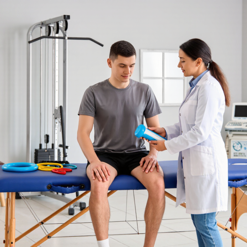
Sports medicine equipment supports three connected tasks: diagnosis, treatment, and prevention. Diagnostic tools may include motion sensors, balance platforms, joint measurement devices, and imaging support systems. They help clinicians observe movement, swelling, strength, and coordination. A careful assessment starts with the athlete’s history, not only a machine reading. Equipment can reveal useful patterns, but it cannot explain every injury alone.
Treatment equipment helps reduce pain, restore movement, and rebuild strength. Examples include therapeutic exercise tools, electrical stimulation units, compression systems, and rehabilitation tables. A clinician may use resistance bands for controlled shoulder work or a balance pad after an ankle sprain. Settings should match the patient’s condition, training stage, and tolerance. More intensity is not always better. Poorly adjusted equipment can increase discomfort or hide warning signs.
Prevention equipment focuses on safer movement and workload control. Wearable monitors can track training volume, while force and flexibility tools may identify uneven loading. Protective supports can improve confidence, but they should not replace proper technique or recovery. Staff should inspect equipment, clean contact surfaces, and record relevant measurements. Even experienced professionals can overvalue precise numbers. Real-world movement is messy. That limitation deserves honest discussion. Combining clinical judgment, current evidence, and patient feedback usually produces a more reliable plan.
Sports medicine equipment helps clinicians connect pain, movement, and tissue damage. MRI systems operating at 1.5–3 tesla can show muscles, tendons, cartilage, ligaments, and bone. A knee scan may reveal a small meniscus tear or hidden swelling. Higher field strength often provides finer detail, but image quality also depends on positioning, timing, and patient movement. MRI does not measure strength or movement directly.
Ultrasound uses high-frequency sound waves to examine soft tissues in real time. Probes ranging from 5–15 MHz can display tendon thickening, fluid, or partial tears near the skin. During an assessment, a clinician may ask the patient to contract a muscle or move a joint. This adds useful functional information. Still, deeper structures may appear less clearly. A bright image can mislead.
Force plates measure ground-reaction forces during walking, jumping, landing, or balance tests. They can reveal uneven loading between the legs, delayed force production, or excessive impact. Numbers alone are not enough. A strong athlete may produce unusual results because of pain, fatigue, or unfamiliar testing conditions. Proper calibration matters. Experienced clinicians compare force data with symptoms, video, strength testing, and training history. Small errors remain possible, especially when the athlete changes technique to avoid discomfort.
Sports medicine equipment turns rehabilitation into measurable practice. A goniometer records joint range of motion, while a hand-held dynamometer measures force during controlled contractions. A balance platform can reveal weight shifts that remain invisible during ordinary walking. These tools help clinicians compare the injured side with the healthy side and track small weekly changes. In practice, I have found that a simple repeatable test often matters more than an expensive device. Consistency is not always easy.
Training load needs a wider view. Session duration, exercise intensity, heart rate, and perceived exertion can be combined into a practical load record. The International Olympic Committee’s 2017 consensus statement recommends monitoring internal and external loads because rapid changes may increase injury risk. However, a single ratio cannot predict every athlete’s response. The body is less tidy than a spreadsheet. The World Health Organization’s 2020 guidelines recommend 150–300 minutes of moderate activity weekly for healthy adults, but recovering athletes may need much less at first. Clinicians should adjust targets according to pain, swelling, sleep, and movement quality. A force test may improve, yet poor balance can still expose a dangerous gap. Repeating ROM, strength, balance, and load measurements under similar conditions creates more reliable evidence for progression. Sources: WHO Guidelines on Physical Activity and Sedentary Behaviour, 2020; IOC consensus statement, British Journal of Sports Medicine, 2017.
Rehabilitation equipment such as digital goniometers and motion-analysis systems helps clinicians measure joint range of motion (ROM) in degrees. The values shown are commonly used adult reference ranges and may vary with age, testing position, and individual health. Other sports medicine equipment can assess strength with dynamometers, balance with force platforms, and training load with wearable motion sensors.
Sports medicine equipment supports the diagnosis and treatment of joint, muscle, tendon, and ligament problems. Arthroscopy equipment is especially useful when a surgeon needs a clear view inside a joint through small openings. A 4-mm arthroscope contains a camera and light system. It can display cartilage, synovial tissue, and damaged structures on a monitor.
Small access matters.
Most procedures use two to four minimally invasive portals. One portal commonly holds the scope, while others allow instruments to enter the joint. Shavers, graspers, probes, and repair tools may pass through these carefully planned pathways. The team chooses each portal according to the joint, injury, and required movement. Knee and shoulder procedures often need different positioning and instrument angles.
A smaller incision can reduce tissue disruption, but it does not remove surgical complexity. The view may become limited when bleeding, swelling, or cloudy fluid affects the image. Good irrigation control and precise camera handling remain essential. Sterile processing also deserves close attention, because delicate optical equipment can lose performance after poor cleaning or damage. In real clinical work, the setup is not always elegant. A portal may need adjustment, and the original plan may change. Training, imaging evidence, patient anatomy, and sound clinical judgment must guide those decisions. Safety should remain more important than speed.
| Equipment or Dimension | Typical Specification | Primary Use | Clinical Function | Key Considerations |
|---|---|---|---|---|
| Arthroscope diameter | Approximately 4 mm | Routine large-joint arthroscopy, including the knee and shoulder | Provides a balance between image quality, irrigation capacity, and access through a small portal | A larger scope generally supports better visualization and fluid flow but may require a slightly larger portal than a smaller-diameter scope |
| Arthroscope viewing angle | Commonly 0° or 30°; other angles may be available | Inspection of joint surfaces, recesses, and compartments | A 0° lens gives a straight-ahead view, while a 30° lens allows the surgeon to look around structures by rotating the scope | The selected angle depends on the joint, target anatomy, and surgeon preference |
| Optical system | Rigid rod-lens or equivalent rigid arthroscopic optical system | Transmission of an illuminated image from inside the joint | Works with a camera head, light source, and monitor to display magnified intra-articular anatomy | Image quality depends on optics, camera resolution, focus, illumination, cleanliness, and correct system connection |
| Camera and display | Camera head connected to a surgical monitor | Real-time visualization during diagnostic and operative procedures | Converts the optical image into a monitor image that can be viewed by the surgical team | White balance, focus, image orientation, and sterile handling should be checked before the procedure |
| Light source and light cable | High-intensity surgical illumination delivered through a compatible cable | Lighting the interior of the joint | Improves visibility of cartilage, synovium, ligaments, menisci, and other intra-articular structures | Light cables and connectors must be compatible with the system; excessive heat at the tip or cable end should be avoided |
| Irrigation system | Sterile fluid inflow with controlled outflow; commonly uses saline solution | Distending and rinsing the joint during arthroscopy | Helps maintain visualization, clears blood and debris, and supports instrument movement | Pressure and flow should be controlled to reduce extravasation and maintain appropriate joint distension |
| Working cannula or sheath | Cannula sized to accommodate the scope or operative instrument | Guiding instruments through a portal | Creates a protected passage for the arthroscope, shaver, probe, graspers, scissors, or other instruments | The internal diameter must match the selected instrument, and the cannula should be positioned to protect surrounding soft tissues |
| Minimally invasive portals | Commonly 2–4 small portals, depending on the joint and procedure | Providing access for visualization, irrigation, and instrumentation | Allows the camera and instruments to enter the joint through separate or shared access points | The number and location of portals vary with anatomy, procedure complexity, instrument trajectory, and the need to avoid neurovascular structures |
| Viewing portal | One portal generally dedicated to the arthroscope | Obtaining a stable visual field | Provides the primary image used to inspect the joint and guide treatment | Portal placement should provide a useful viewing angle and permit adequate visualization of the operative area |
| Working portal | One or more portals for operative instruments | Performing treatment under direct visualization | Allows passage of probes, graspers, punches, shavers, radiofrequency devices, or suture instruments | Instrument trajectory, triangulation, portal spacing, and soft-tissue protection are important for precise handling |
| Diagnostic arthroscopy | 4-mm scope with appropriate viewing angle and irrigation | Evaluating internal joint structures | Assesses cartilage, synovium, menisci, labrum, ligaments, and other accessible structures for injury or disease | Arthroscopy is an invasive procedure and should be performed by appropriately trained clinicians using sterile technique |
| Operative arthroscopy | Scope plus compatible instruments through 2–4 portals | Minimally invasive treatment of selected joint conditions | Supports procedures such as tissue trimming, loose-body removal, synovial treatment, meniscal work, and selected ligament or cartilage procedures | The equipment configuration must be selected for the specific procedure and should include appropriate backup instruments |
| Shaver system | Powered rotating or oscillating blade with suction | Removing or contouring soft tissue and selected damaged tissue | Combines mechanical tissue removal with fluid evacuation to help maintain the operative field | Blade selection, suction level, and activation technique should be controlled to avoid unintended tissue injury |
| Probe and grasping instruments | Handheld instruments passed through a working cannula | Palpating, stabilizing, retrieving, or manipulating tissue | Helps assess tissue integrity and handle structures during diagnosis or repair | Instrument size and jaw design should correspond to the joint space and intended task |
| Sterilization and reprocessing | Validated cleaning, disinfection, and sterilization process appropriate to the device | Preparing reusable equipment for safe clinical use | Reduces the risk of cross-contamination and preserves device function | Manufacturer instructions, facility protocols, inspection, leak testing, and correct packaging must be followed for reusable equipment |
| Main benefits | Small incisions, direct visualization, and targeted instrumentation | Minimally invasive joint evaluation and treatment | May reduce soft-tissue disruption compared with open approaches and can support accurate intra-articular assessment | Patient outcomes depend on the diagnosis, procedure, clinician expertise, sterile practice, and postoperative care—not equipment alone |
Note: Specifications are general clinical ranges and may vary according to joint, procedure, patient anatomy, instrument design, and applicable medical-device regulations.
Sports medicine equipment includes braces, rehabilitation systems, diagnostic tools, and therapeutic devices. Their safety depends on more than comfort or appearance. The FDA groups medical devices into three classes according to risk and required controls.
FDA classification data commonly shows about 47% of devices as Class I, 43% as Class II, and 10% as Class III. Class I devices usually face general controls. Class II products may require special controls or 510(k) clearance. Class III devices face the highest risk and often need premarket approval.
That distinction matters. A powered rehabilitation device may look simple, yet software faults, overheating, or incorrect force can cause injury. The FDA’s device databases and recall reports show why manufacturers must monitor products after release. Not always.
ISO 13485 adds a quality framework across the product life cycle. The ISO Survey reports tens of thousands of ISO 13485 certificates worldwide, reflecting broad adoption of controlled medical-device processes.
These controls cover design verification, risk management, supplier evaluation, traceability, complaint handling, and corrective action.
In practice, a manufacturer should document how a knee-support sensor performs under repeated movement, sweat, cleaning, and impact. Certification is not a safety guarantee. It can still expose weak assumptions. A rushed risk review, incomplete user testing, or poorly documented software change may escape attention. Clinicians and buyers should check intended use, warnings, maintenance instructions, and evidence supporting performance before selecting equipment.
It supports diagnosis, treatment, rehabilitation, and injury prevention. Tools can measure movement, balance, swelling, strength, and coordination. A machine cannot explain every injury alone.
Motion sensors, balance platforms, joint measurement devices, and imaging support systems can reveal useful patterns. Clinicians should also review injury history and physical findings. Numbers can mislead.
Resistance bands can support controlled shoulder exercises. Balance pads may help after an ankle sprain. Electrical stimulation, compression systems, and rehabilitation tables can support recovery. Settings should match tolerance and training stage.
Not always. Excessive force or stimulation may increase discomfort or hide warning signs. The clinician should adjust settings after observing pain, movement, and fatigue. More is not automatically better.
Wearable monitors can track training volume. Force and flexibility tools may reveal uneven loading. Protective supports can improve confidence, but technique and recovery still matter. Real movement remains messy.
An arthroscope uses a small camera and light to display structures inside a joint. Surgeons may view cartilage, synovial tissue, and damaged areas. The image can become unclear because of bleeding, swelling, or cloudy fluid.
Many procedures use two to four small portals. One portal may hold the camera, while others admit probes, graspers, shavers, or repair tools. Portal placement depends on the joint and injury.
Powered devices can overheat, apply incorrect force, or develop software faults. Quality controls should cover testing, risk review, supplier checks, traceability, maintenance, and complaints. Certification helps, but it is not a complete safety guarantee.
They should review intended use, warnings, cleaning instructions, maintenance needs, and performance evidence. Staff should inspect devices and clean contact surfaces. A rushed review may miss a serious weakness.
Sports Medicine Equipment includes the tools used to diagnose, treat, and prevent athletic injuries throughout the care process. Diagnostic systems may include 1.5–3 T MRI scanners for detailed soft-tissue and bone imaging, 5–15 MHz ultrasound devices for real-time assessment of muscles and tendons, and force plates that measure loading patterns, balance, and movement mechanics. Together, these technologies help clinicians identify problems accurately and design individualized treatment plans.
Rehabilitation equipment supports the measurement and improvement of range of motion, muscle strength, balance, coordination, and training load. Arthroscopy equipment, such as 4-mm scopes used through two to four minimally invasive portals, can assist with internal joint examination and selected procedures. Because these devices directly influence patient care, safety is essential. The FDA’s three-class risk system guides regulatory oversight, while ISO 13485 quality controls promote consistent design, manufacturing, testing, and documentation.